|
|
PSY 340 Brain and Behavior Class 36: Lateralization of Function |
|
|
|
PSY 340 Brain and Behavior Class 36: Lateralization of Function |
|
A. The Right and Left Hemispheres
Each side of the brain (the right and the left hemispheres) has its own work to do: processing incoming stimuli and sending out motor instructions. The work of the brain is divided between the hemispheres which communicate (as we will see below) with each other across the corpus callosum.
Lateralization: Refers to those behaviors and cognitive abilities that each hemisphere specializes in. For example, language ability is primarily localized in the left hemisphere.
Contralateral = the other side while ipsilateral = the same side
Function
Left Hemisphere
Right Hemisphere
Muscles
Contralateral (right side)
Contralateral (left side)
Skin Receptors
Contralateral (right side)
Contralateral (left side)
Vision (Eyes)
Contralateral (Right visual field of both left and right eyes; see diagram below)
Contralateral (Left visual field of both left and right eyes; see diagram below) Hearing (Ears)
Information from both ears, but stronger from right ear
Information from both ears, but stronger from left ear
Taste
Ipsilateral (left side of tongue)
Ispilateral (right side of tongue)
Smell
Ipsilateral (left nostril)
Ipsilateral (right side of tongue)
Trunk Muscles & Facial Muscles
Jointly controlled
B. Visual and Auditory Connections
Visual Processing. Our eyes are connected so that visual information from the right and left visual fields are processed on the contralateral side of the brain (see diagram below) with half the optic nerve crossing over at the optic chiasm.
- Right visual field
left half of each retina
- Left visual field
Auditory Processing. Information from each ear is transmitted to both sides of the brain. However, each hemisphere processes the information from the opposite each with greater attention.
- The reason why each hemisphere receives information from both ears arises from the need to detect where sound is coming from in the outside environment. The brain can compare tiny differences between the information from each ear (e.g., timing or volume differences) in order to figure out where the sound comes from.
C. Corpus Callosum and the Split Brain Operation
The corpus callosum is a massive set of axons which allow the two hemispheres to exchange information with one another. There are an estimated 200 million axons which cross from one side of the brain to the other in this "commissure" (a brain "commissure" is a a collection of axons which link brain areas in each hemisphere).
Research has shown that a significant proportion of the axons in the corpus callosum (perhaps 1/3rd) are inhibitory, that is, they inhibit activity in the other hemisphere of the brain.
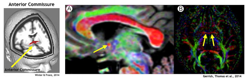
There are two other significant commissures: the anterior commissure (located below the corpus callosum near the front of the brain; contains about 3.5 million axons) and the hippocampal commissure (located below the corpus callosum near the rear of the brain). These commissures are seen in a diagram on the right. (Note that there are also two other very small commissures: posterior & habenular)
1. Severing the corpus callosum prevents the sharing of most information between the brain hemispheres.
2. Epilepsy = Condition characterized by repeated episodes of excessive synchronized neural activity (i.e., seizure).
The causes of epilepsy are many including brain trauma, infection, and genetic abnormalities. Most frequently, though, we do no know why someone has epilepsy. Roughly 1-2 % of the population experiences epilepsy.
YouTube Video. Video of a grand mal (tonic-clonic) seizure (1'18") of a college-age man, Josh. (Note that this is a graphic video.)
Since epilepsy represents an increased tendency of neurons to fire (i.e., they tend to be hyperpolarized), general treatment of epilepsy uses drugs which increase GABA functioning (i.e., are inhibiting). Most people with epilepsy (90%) can control their condition with medications that suppress seizure activity. Surgery for epilepsy can take two forms. The earliest form sought to remove any focus of epileptic activity, that is, a "focus" is a localized site of scar tissue, often on the surface of the cortex, that promotes seizures.
3. Commissurotomy (also called corpus callosotomy). If seizure activity is not controlled by drug therapy or focus removal, a small number of people experience repeated and life-threatening seizures. Thus, some epileptic patients have their corpus callosum severed to prevent seizure activity from crossing from one hemisphere to the other. As Luat et al. (2017) state, "the corpus callosum is the major pathway for the interhemispheric spread of
[electrical-epileptic] discharges and its disconnection leads to a disruption of rapid seizure spread" (p. 624). The pathway for most epileptic seizure activity comes in the anterior portion of the corpus callosum, i.e, the genu. These individuals are often referred to as split-brain people. Research has found that this operation has good to excellent results particularly in patients whose epilepsy caused them to fall to the floor ("drop seizures") as well improving the quality of life (Untenberger et al., 2016). Note that the surgery is often performed in two stages: in the first stage, only the front 2/3rds of the corpus callosum is severed (leaving intact the visual system's interhemispheric connections). If the 1st stage does not achieve the desired clinical reduction in seizure activity, a 2nd stage operation completes the severing of the remaining corpus callosum.
Such operations began in Los Angeles 1962 and built upon work done in the early 1950s by Roger W. Sperry (1913-1994) who researched the effect of severing the corpus callosum in cats. He and Michael Gazzaniga began to study the effects of split-brain operations on patients and what they revealed about the functioning of each hemisphere of the brain. Sperry won the Nobel Prize in Medicine & Physiology in 1981 "for his discoveries concerning the functional specialization of the cerebral hemispheres" as a result of this work.
4. Studying Split-Brain Patients. What did Sperry and Gazzaniga do to study split-brain patients? Information is shown in either the left or right visual fields and, thus, is transmitted only to the opposite hemisphere of the brain which processes that visual field. So, as shown in the figure, if a telephone is shown in the right visual field and, thus, processed in the left hemisphere, the patient will normally say that they saw a telephone. But, if information is projected in the left visual field and processed in the right hemisphere, the patient will report that they saw nothing. Why? Speech is processed in the left hemisphere for most people and, thus, the patient's speech can only truthfully report that nothing was presented.
However, if a word like "Dog" is presented to the right hemisphere via the left visual field, the individual will not report seeing anything. However, if they are asked to use their left hand to point to what they might have seen (or to draw something), the split-brain patient will point to or draw a dog with the left hand (which, of course, is under the control of the right hemisphere).
YouTube Video: Split-brain patient 'Joe' being tested with stimuli presented in different visual fields (10'11"). In a 1996 science special on PBS, Alan Alda visits with Mike Gazzaniga at Darmouth College (he's now at the University of California, Santa Barbara). They are joined by Joe, a "split-brain" person with whom Mike has worked for many years. In this clip Michael Gazzaniga demonstrates how split-brain patients are tested. Often because the left hemisphere (which has language) isn't aware of what the right hemisphere saw and did, the left hemisphere has to make up a story to explain that behavior.
Developed by Gazzaniga and his doctoral student, Joseph LeDoux, this is a theory of the "left hemisphere interpreter" = the tendency of the left hemisphere to invent explanations for behaviors which arose from unconscious sources or for experiences in the world which don't make complete sense. He explains this theory in his 2011 book, Who's In Charge?
- The right hemisphere is much more tied to what is concrete and "real".
5. The two hemispheres of a split-brain person process information independently of each other. However, the brain eventually learns to use smaller connections between the left and right hemispheres to avoid conflicts between them.
6. The Right Hemisphere vs. the Left Hemisphere: A Preliminary Overview
a. The right hemisphere is better than the left at perceiving the emotions in people's gestures (non-verbal or paralinguistic information).
b. People with right hemisphere damage speak with less inflection and expression, plus they often have trouble interpreting the emotions that other people express through their tone of voice (prosody).
c. Research findings suggest that the right hemisphere is more adept than the left at comprehending spatial relationships.
d. The left hemisphere is more focused on details and the right hemisphere is better at perceiving overall patterns.
NOTE: In the next class we will examine the claims by the British neuroscientist and psychiatrist, Iain McGilchrist that there are greater differences between the right and left hemisphere than you see below in the table summarizing our textbook.
Role of Left Hemisphere Role of Right Hemisphere Speech Production of speech, comprehension of the literal meaning of speech Emotional inflections, understanding jokes & humor, sarcasm, emotional content of speech Auditory System Sounds related to speech Non-language environmental sounds (e.g., rain)
MusicEmotions Expressions of happiness and anger (= denial of happiness)
Expressions of fear, disgust, sadness; interpreting the emotional expressions of other people Vision Details Overall configuration;
spatial processing (e.g., arranging pieces of a puzzle or drawing a picture)Mode or Style
(How data are processed)Details, parts, pieces Gestalt, overall configuration; global form
D. Development of Lateralization and Handedness
1. Planum temporale: A section of the temporal cortex that is larger in the left hemisphere in approximately 65% of the population. This difference in size is apparent at age 3 months in humans. Children with the biggest ratio of left to right planum temporale performed best on language tests. Here is another example of hemispheric asymmetry.
2. Corpus Callosum. The corpus callosum matures slowly over the first 5 to 10 years of human life. Because the neurons connected by the corpus callosum take years to develop their mature adult pattern, the behavior of young children sometimes resembles that of split-brain people.
3. Agenesis of the Corpus Callosum (ACC). People born without any or a partially-formed corpus callosum (ACC) can perform some tasks that split-brain people fail, possibly due to larger-than-normal hemispheric connections developing elsewhere in the brain. For example, they can describe what they feel with either hand and what they see in either visual field.
The following two commissures are often larger than normal in people born without a corpus callosum:
a. Anterior commissure (in image A above, the yellow arrow points to a very small red dot which is the anterior commissure): Connects the two hemispheres around the anterior parts of the cerebral cortex. It seems to be mostly involved in smell in neurologically intact persons. It also seems to convey visual information in ACC patients.
b. Hippocampal commissure: Connects the left hippocampus to the right hippocampus. Very little is know about the function of this connection. There is some evidence that it is involved in recognition memory, that is, knowing that you have seen words or images before rather than for the first time.
A study by Paul et al. (2003) came to this conclusion about language deficits in individuals with ACC:
"...normally intelligent individuals with ACC are impaired in the understanding of non-literal language and emotional-prosodic cues that are important in social communication. .. the performance of individuals with ACC was similar to patients with right hemisphere brain damage. Thus, persons with ACC appear to lack interhemispheric integration of critical aspects of language processed by the right hemisphere." (Paul et al., 2003, Abstract)
Handedness
- Roughly, 90% of population is right-handed and 10% either left-handed or ambidextrous.
- General estimate of left hemisphere dominance for speech: roughly 95% of right-handers & 80% of left-handers.
- Many left-handers also show > than normal spatial processing on the left (not right) side.
Right-handers have preference for turning left and left-handers a preference for turning right when confronted with forks in the road.
As we will see, McGilchrist links the left hemisphere with seeking out prey which is usually caught/grasped by the right hand.
Avoiding Overstatements
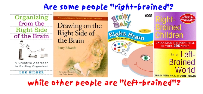
What is the answer to the first question at the beginning of this lecture? Are there "right-brained" and "left-brained" people? NO: This is a myth!
As our text points out, such claims are based on two correct and one incorrect assumptions:
- each hemisphere specializes in particular functions [TRUE]
- tasks involving particular functions evoke higher levels of activity in particular hemispheres [TRUE]
- individual persons rely more heavily and most of the time on one of the hemispheres [FALSE]
We do not have scientific evidence to support that last claim: that people rely upon either the right or the left hemisphere habitually.
- Indeed, the presence of the corpus callosum means that the brain is constantly engaged in interhemispheric communication: one side of the brain is always in conversation with the other side of the brain.
- Most cognitive processes (including creativity, imagination, seeing details, doing mathematics, understanding language, etc.) actually rely upon functions of both hemispheres.
However, we will see more about right versus left brain functioning in the next lesson on the work
of Dr. Iain McGilchrist and this theory of The Master and the Emissary.
References
Andermann, R., & Hart, Y. (1999). Rasmussen's syndrome. Brussels, Belgium: International League against Epilepsy. Retrieved 4/20/05 from the Web site: http://www.epilepsy.org/ctf/rasmussens_syndrome.html
Bien, C. G., Widman, G., Urbach, H., Sassen, R., Kuczaty, S., Wiestler, O. D., Schramm, J., & Elger, C. E. (2002). The natural history of Rasmussen's encephalitis. Brain, 125(8) 1751-1759. https://doi.org/10.1093/brain/awf176
Banich, M. T. (1997). Neuropsychology: The neural basis of mental function. Boston: Houghton-Mifflin.
Cohen, I., David, E., & Netanyahu, N. S. (2019). Supervised and Unsupervised End-to-End Deep Learning for Gene Ontology Classification of Neural In Situ Hybridization Image. Entropy, 21. https://dx.doi.org/10.3390/e21030221
Funnell, M. G., Corballis, P. M., & Gazzaniga, M. S. (2000). Cortical and subcortical interhemispheric interactions following partial and complete callosotomy. Archives of Neurology, 57, 185-189. https://doi.org/10.1001/archneur.57.2.185
Gerrish, A. C., Thomas, A. G., & Dineen, R. A. (2014). Brain white matter tracts: Functional anatomy and clinical relevance. Seminars in Ultrasound, CT, and MRI, 35(5), 432-444.
https://doi.org/10.1053/j.sult.2014.06.003Hofer, S., Karaus, A., & Frahm, J. (2010, April 13). Reconstruction and dissection of the entire human visual pathway using diffusion tensor MRI. Frontiers in Neuroanatomy. doi: 10.3389/fnana.2010.00015
Kolb, B., & Whishaw, I. Q. (2003). Fundamentals of human neuropsychology (5th ed.). New York: Worth Publishing
Luat, A. F., Asano, E., Kumar, A., Chugani, H. T., & Sood, S. (2017). Corpus callostomy for intractable epilepsy revisited: The Children's Hospital of Michigan series. Journal of Child Neurology, 32(7), 624-629. https://doi.org/10.1177/0883073817697847
Luck, S. J., Hillyard, S. A., Mangun, G. R., & Gazzaniga, M. S. (1989, November 30). Independent attentional systems mediate visual search in split-brain patients. Nature, 342, 543–545. https://doi.org/10.1038/342543a0
McGilchrist, Iain. (2021). The matter with things: Our brains, our delusions and the unmaking of the world. London, UK: Perspectiva Press.
McManus, C. (2002). Right hand, left hand: The origins of asymmetry in brains, bodies, atoms, and cultures. Weidenfeld & Nicolson.NINDS. Agenesis of the corpus callosum information page. Bethesda, MD: National Institute of Neurological Disorders and Stroke. http://www.ninds.nih.gov/disorders/agenesis/agenesis.htm
Paul, L. K., Van Lancker-Sidtis, D., Schieffer, B., Dietrich, R., & Brown, W. S. (2003). Communicative deficits in agenesis of the corpus callosum: Nonliteral language and affective prosody. Brain and Language, 85(2), 313-324. https://doi.org/10.1016/S0093-934X(03)00062-2
Szaflarski, J. P., Binder, J. R., Possing, E. T., McKiernan, K. A., Ward, B. D., & Hammeke, T. A. (2002). Language lateralization in left-handed and ambidextrous people: fMRI data. Neurology, 59(2), 238-244. https://doi.org/10.1212/WNL.59.2.238
Unterberger, I., Bauer, R., Walser, G., & Bauer, G. (2016). Corpus callosum and epilepsies. Seizure, 37, 55-60. https://doi.org/10.1016/j.seizure.2016.02.012
Zimmer, C. (2009, May). The brain: The big similarities & quirky differences between our left and right brains. Discover Magazine. Accessed 4/16/09 from the website: http://discovermagazine.com/2009/may/15-big-similarities-and-quirky-differences-between-our-left-and-right-brains
The first version of this page was posted on April 20, 2005
![[Brain Image]](../graphics/head_space.gif)
![[Visual System connections]](../graphics/visualsystem2.png)