![[Brain Image]](../graphics/head_space.gif)
PSY 340 Brain and Behavior
Class 19: How the Brain Processes Visual Information/Early Experience OUTLINE
|
|
PSY 340 Brain and Behavior Class 19: How the Brain Processes Visual Information/Early Experience OUTLINE |
|
This outline page is meant to be used in class to accompany
- the longer notes and summary diagram (in pdf format) that I posted on the Lectures page.
- You may bring a copy of the summary diagram with you to Test #2 in addition to the one-sided cheat sheet page.
I am combining Sections 5.2 & 5.3 in our textbook and treating them according to my own approach.
![[Diagram of Eye and Retinal Layers]](../graphics/eye_retina.jpg)
Bipolar Layer: Lateral Inhibition
![[Lateral Inhibition]](../graphics/Lateral%20Inhibition2.png)
Ganglion Layer: The Notion of Receptive Fields
The ganglion layer contains between 18-20 different types of ganglion cells. For our purposes, the three most important involve:
- Parvocellular (parvo = small) (or midget ganglion or X ganglion) responsive to detail, color
- Magnocellular (magno = large) (or parasol ganglion or Y ganglion) responsive to movement, overall pattern, no color
- W ganglion: project to the koniocellular cells (K-cells) of the lateral geniculate nucleus. Functions are unclear.
- When we discuss sleeping and waking, we will look at a 4th type: intrinsically photosensitive ganglion cells.
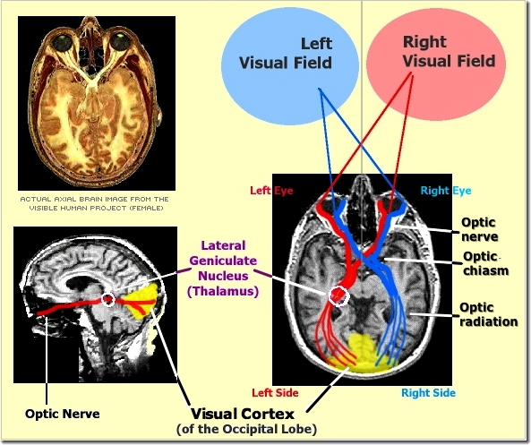
Pathway from Retina to Occipital Lobe

Summary Diagram
Occipital Cortex: Blindsight & Processing
Damage to occipital cortex leads to blindness, but some people experience blindsight = YouTube Video (38 secs.)
Why? How can this be?
1. There may be small areas of healthy tissue: not enough for conscious perception, but enough to support some functions
2. Possibly because of connection between koniocellular layer of the LGN and area MT/V5 (Ajina et al. 2015; see below)
Processing Shape: Occipital Cortex
Hubel & Wiesel's Cat Experiment: Visual Cortex Response to Light
• First at Johns Hopkins and then Harvard University Medical School David Hubel and Torsten Wiesel worked as researchers on the visual system. They recorded electrical activity from individual neurons in the brains of cats. Their findings won them the Nobel Prize for Physiology or Medicine in 1981.
• Video of recording from visual cortex cell recording (3'15")
Occipital cortex is extracting shape information from data in V1 & V2
- Areas V1 (primary visual cortex) & V2 (secondary visual cortex)
- Simple Cells (V1 only): fixed excitatory or inhibitory zones. Responsive to bars of light in vertical, horizontal, or intermediate orientations.
- Complex Cells (V1 & V2): responds mostly to patterns of light oriented in a specific way within a relatively large receptive field, e.g., bar at 45 degree angle moving.
- End-stopped (or “Hypercomplex”) Cells (V1 & V2): like complex cells, but also highly responsive to bars with end-points. Very large receptive fields.
- Areas V1/V2 connect to the inferior temporal cortex (in the “ventral stream”) which responds to very complex shapes in three-dimensional space. These neurons contribute toward the experience of shape constancy
Stereoscopic Depth Perception
- Initial experience arises in V1 since data from each eye come together in this region
- Additional processing probably in V3
The Dorsal vs. Ventral Streams
Dorsal (Where/How) Stream [Parietal Lobe]
- Knowing location of objects in space
- Linking visual data with movement & action
- New (2017) research: responsive to overall shape in order to allow movement/action to be successful, but NOT enough to identify object
Ventral (What) Stream (What/Object Recognition) [Temporal Lobe]
- Oriented toward the perception of visual information and location of multiple disorders of object recognition
- Color Constancy: V4 (Temporal-parietal junction) [Land's Retinex Theory of Color Processing]
- Visual Motion Detection: Middle Temporal Cortex (V5) & Middle Superior Temporal Cortex [MST]
- Motion of objects is not confused by any motion of the head. We can keep these two types of motion separate in our vision.
- Damage: ? motion blindness
Parahippocampal Place Area: Recognize physical environment & geography
- Damage leads to difficulty or inability to learn to navigate an environment or recognize where one is located
- Visual Word Form Area (VWFA): LEFT lateral occipito-temporal sulcus
- Decoding letters & words
- Damage leads to pure alexia = inability to read words
- Face Recognition: Fusiform gyrus (larger in Right hemisphere)
- Damage leads to prosopagnosia = inability to recognize the faces of other people
- Visual agnosia = inability to recognize objects despite satisfactory vision. Usually due to damage of the temporal lobe.
- Shape & Object Recognition: ITC (inferior temporal cortex)
A. Infants & Vision
- Newborn Infants have a preference for faces [real or distorted] as long as eyes are on the top.
- Inability to look away from moving objects to stationary objects (even still difficulty at 6-7 years old)
B. Early Experience: Impact upon Visual Development
- Experiences "fine tune" the neural visual system to see the world accurately
- Sensitive or critical periods for experiences to have effect upon the nervous system
- Strabismus (eyes not point in same direction) ==> impaired stereoscopic depth perception
- Amblyopia ("lazy eye") can lead to blindness in eye. Eye patch used to treat it
- Impaired vision in infancy and early childhood leads to long-term visual defects in adulthood
- Cataracts corrected after age 7: children have impairment for motion & depth perception
The first version of this page was posted on March 7, 2007