Feb 9, 2005
![[Brain Image]](../graphics/head_space.gif)
PSY 340 Brain and Behavior
Class 13: Research Methods (OUTLINE)
|
Feb 9, 2005 |
PSY 340 Brain and Behavior Class 13: Research Methods (OUTLINE) |
|


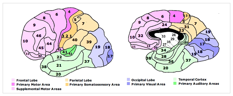
How do we learn about how the brain works?
1. Look at the Effects of Brain Damage
2. Stimulate Some Brain Area and Analyze the Resulting Behavioral Change
3. Correlate Brain Anatomy with Behavior
4. Record Brain Activity during Behavior
1. Look at the Effects of Brain Damage
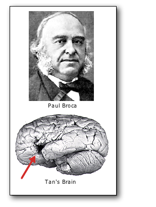

• Paul Pierre Broca (1824-1880)
• Animal Brain Research
- Lesion (damage tissue) or
- ablation (tissue removal)
- Insert electrode in animal's brain via stereotaxic instrument
NOTE: Until the advent of modern technologies [described below] in the late 20th and early 21st centuries, it was not possible to examine directly brain damage in a living human being. As Broca's work showed, brain damage could only be seen at autopsy after the death of a patient.
• Transcranial Magnetic Stimulation (TMS): A non-invasive technique that passes a focused, intense, and changing magnetic field though the scalp and skull
- Can cause both stimulation and a temporary functional stoppage of neural tranmission (i.e., "temporary damage" without permanent damage"
- BBC The Brain: A Secret History Michael Mosley has areas of his brain turned off by TMS (YouTube)
2. Stimulate Some Brain Area and Analyze the Resulting Behavioral Change
• Originally, electrical stimuli applied to the brain to see the resulting behavior
Optogenetics = Using light to control a limited group of neurons
- "Optogenetics is the combination of genetic and optical methods to cause or inhibit well defined events in specific cells of living tissue and behaving animals" (Deisseroth, 2015, p. 1213). This technique was pioneered by Keith Deisseroth of Stanford University beginning about 2005.
- A specially-designed virus is used to insert a light-sensitive protein into the membrane of a particular type of neuron in the brain of a living animal
- NATURE METHODS: Method of the Year 2010: Optogenetics Video (YouTube) • "This video shows how scientists can control the behaviour of cells simply by switching on a light."
3. Correlate Brain Anatomy with Behavior
- Computerized Axial Tomography (CAT): Brief x-rays from 180 degrees around head. First used in the 1970s.
- Magnetic Resonance Imaging (MRI): Body tissue is subjected to a strong magnetic field which is turned on and off rapidly in the presence of a radio wave. The atoms of the brain change their alignment (spin) because of the magnetic field when it is on and give off characteristic radio signal when it is turned off. A detector reads those signals and, using a computer, can map the structure of the tissue. Developed in the 1970s and used clinically beginning in the 1980s.
In the MRI image on the right, the patient appears to have a large tumor growing in the mid-portion of the brain. This has compressed the cingulate cortex and is pressing down on the corpus callosum.
4. Record Brain Activity during Behavior
• EEG (Electroencephalograph): Measuring brain waves via skull electrodes. First developed in 1924, 100 years ago.

• PET Scan (Positron Emission Tomography)
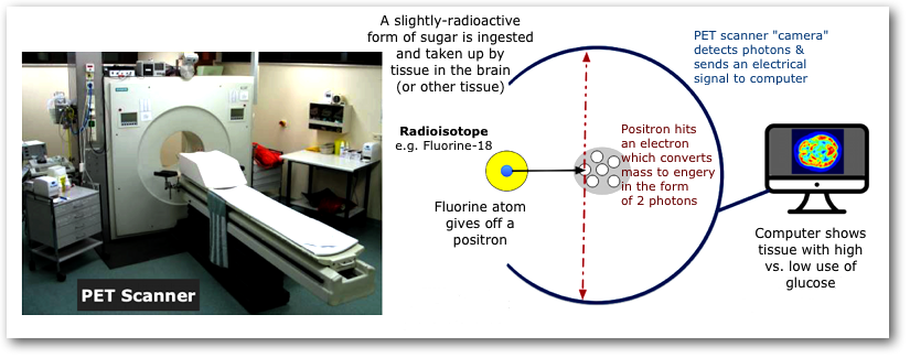
• Functional Magnetic Resonance Imaging (fMRI)
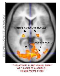
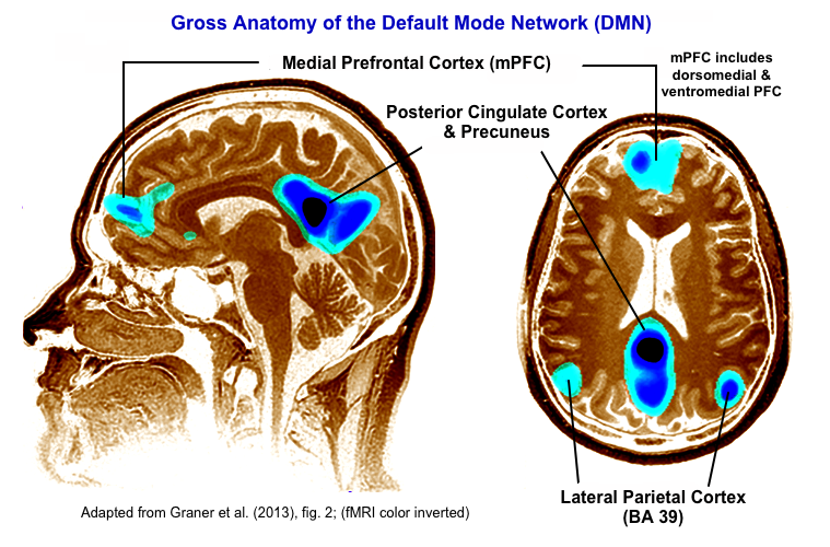
- Functioning or activity
- BOLD (= Blood Oxygen Level Dependent) signal
- Default Mode Network (DMN): What are you doing when you are daydreaming? Imagining the future? Reminiscing about past experiences? Results from fMRI studies show that there is a specific network of brain areas which are ACTIVE when your mind is wandering in these ways. We'll see more in the next class.
- Problem of head movement
- Limited resolution scale
Emerging Techniques. Note that there are a host of new techniques of imaging or studying the brain which we will not review and which you will not be responsible for knowing. These include CLARITY Imaging of Tissue (Postmortem), Magnetoencephalography, Magnetic Resonance Spectroscopy, Near Infrared Spectroscopy, Syringe-Injectable Electronics, and Ultrasound Neuroimaging. For anyone in the class who expects to enter a career in neuroscience or neuro-related medicine and health care, these kinds of techniques will doubtless become more and more important in the decades ahead.
Brain Size and Intelligence
- Humans do not have the largest brains across animal species
- Humans: Brains vs. IQ (Intelligence)
- There is a moderate correlation between brain size and IQ tests (r = ~0.30-0.40, that is, 9-16% of IQ scores related to brain size).
- CAUTION: IQ test scores may not be adequate to evaluate "intelligence" which is a notoriously difficult concept to quantify.
- Hevern: Intelligence involves those abilities to cope successfully within whatever environments (physical, interpersonal, or cultural) individuals find themselves.
- Males generally do have larger brains than females (roughly 8-10%). However, there are no overall IQ differences between men and women.
- Males vs. Females
- Women have deeper & more sulci than men and, despite females brains being roughly 8-10% smaller in volume, there is equal cortical surface for men and women.
- Some structural differences have been found in the wiring between female and male brains. The significance of these findings is not clear.
- Structural differences ≠ behavioral differences, for example, Males > Females in face processing cortical area, yet, Females > Males in actual face processing tasks
- Is there a sex/gender (s/g) difference in the human brain? Except for an overall size difference as noted above, comprehensive meta-analyses of sex/gender brain differences and their relationship to actual behavior shows very little difference
This page was first posted February 3, 2005