February 13, 2025
![[Brain Image]](../graphics/head_space.gif)
PSY 340 Brain and Behavior
Class 13: Research Methods in Neuropsychology
|
February 13, 2025 |
PSY 340 Brain and Behavior Class 13: Research Methods in Neuropsychology |
|
Note: The material in this class wlll not appear on Test #1, but on Test #2.
The Problem Solved by Korbinian Brodmann (1868-1918)
The rapid development of neurology and psychiatry in the second half of the 19th and early part of the 20th century presented researchers with a significant problem in communication. There were agreed-upon terms for the major lobes and gyri of the human cerebral cortex. However, there remained a difficulty in identifying subregions of the cortex in a way which would be understood by scientists in different nations who spoke different languages. The work of Korbinian Brodmann helped to settle that problem.
Brodmann examined the cellular and laminar structure of the human cortex and the cortical tissue of other animals. Eventually he published his important monograph on the cytoarchitectonic structure of the human cortex in 1909. "Cytoarchitectonic" means the architecture of the cells (cyto = cell in Greek) Dr. Laurence Garey (1994) notes:
The basis of Brodmann's cortical localisation is its subdivision into 'areas' with similar cellular and laminar structure. He compared localisation in the human cortex with that in a number of other mammals, including primates, rodents and marsupials. In humans, he distinguished 47 areas, each carrying an individual number, and some being further subdivided.
Brodmann's numbering of these cortical locations has become one of the standard ways in which clinician identify brain areas. These are generally known as "Brodmann Areas" (BA) and will often be cited in texts, for example, as "in BA 45 and 46...". It is presumed that the informed reader will know already or have access to a map of these areas (see below):
How do we learn about how the brain works?
In one of my classes a while ago, a student asked, "So, how do we know these things about the brain?" Indeed, how do we know that the occipital lobe deals with vision and the frontal lobe with working memory? Why can we say that the hippocampus is responsible for any new declarative memories, but NOT for procedural memories or learning like how to ride a bike?
1. Look at the Effects of Brain Damage
2. Stimulate Some Brain Area and Analyze the Resulting Behavioral Change
3. Correlate Brain Anatomy with Behavior
4. Record Brain Activity during Behavior
1. Look at the Effects of Brain Damage
Paul Pierre Broca (1824-1880)
- Patient could only say, "tan, tan" after stroke.
- Autopsy of brain (see left) found damage to lateral posterior (side, rear area) of frontal lobe.
- Broca's or expressive aphasia: inability or difficulty speaking clear language.
Animal Brain Research
- Done most frequently with rats & mice, sometimes rabbits, and occasionally monkeys
- Cause either a lesion (damage) or ablation (tissue removal) in the animal's brain & observe behavior afterward
- Insert electrode in animal's brain via stereotaxic instrument
NOTE: Until the advent of modern technologies [described below] in the late 20th and early 21st centuries, it was not possible to examine directly brain damage in a living human being. As Broca's work showed, brain damage could only be seen at autopsy after the death of a patient.
Transcranial Magnetic Stimulation (TMS)
- For both treatment and research purposes, transcranial magnetic stimulation (TMS, see above) is an emerging, non-invasive method by which to affect underlying brain tissue by passing a focused, intense, and changing magnetic field though the scalp and skull. Recall the principle of electromagnetic theory that anytime an electrical current passes through a wire, it causes a magnetic field to generate at a 90º angle (perpendicular to the wire).
- TMS can cause brain tissue either to be stimulated (depolarizing neurons) or to stop functioning temporarily (hyperpolarizing neurons). Thus researchers can investigate the relationship between precise areas of the brain's tissue and the resulting impairment without causing long-term damage to the brain
- From BBC The Brain: A Secret History: Michael Mosley has areas of his brain turned off by TMS (YouTube)
2. Stimulate Some Brain Area and Analyze the Resulting Behavioral ChangeIn the 19th & early 20th centuries, physiologists began to stimulate the brains of experimental animals (dogs in particular) by using an electrical stimulus. This research established that some important areas of the brain were responsible for certain sensory experiences and motor skills.
Optogenetics = Using light to control a limited group of neurons
- "Optogenetics is the combination of genetic and optical methods to cause or inhibit well defined events in specific cells of living tissue and behaving animals" (Deisseroth, 2015, p. 1213). This technique was pioneered by Keith Deisseroth of Stanford University beginning about 2005.
- A specially-designed virus is used to insert a light-sensitive protein into the membrane of a particular type of neuron in the brain of a living animal
- When a very thin optical light shines on those neurons, it causes the neurons either to turn on (excitatory response by opening a + sodium channel) or to turn off (inhibitory response by opening a - chlorine channel). The researcher can then see what changes in behavior take place.
3. Correlate Brain Anatomy with Behavior
Computerized Tomography (CT) (formerly called Computerized Axial Tomography (CAT)
- This diagnostic and research technique was developed in the 1970s to provide images of the inside of a person's body. It is primarily used as a diagnostic tool in medicine.
- Even though x-rays generally do not show much variation in soft tissues like the brain, multiple brief x-rays which are taken around the head can be processed by a computer to detect very small variations as the x-rays pass through even soft tissue. When combined by the computer, this technique can generation a picture of what the underlying tissue looks like.
Images will show areas of damage. For example, in the CAT scan image on the right, the individual has experienced a cerebral hemorrhage (a type of stroke). Indeed, in most emergency rooms, if a patient is brought in with a suspected stroke or other brain injury, the first type of imaging they will receive is a CT scan.
Magnetic Resonance Imaging (MRI)
- This type of imaging of the inside of the living human body was developed in the 1970s and 1980s and is used primarily as a diagnostic tool in medicine.
- Body tissue is subjected to a strong magnetic field which is turned on and off rapidly in the presence of a radio wave. The atoms of the brain change their alignment (spin) because of the magnetic field when it is on and give off characteristic radio signal when it is turned off. A detector reads those signals and, using a computer, can map the structure of the tissue. There is NO radioactive materials used in an MRI.
In the MRI image on the right, the patient appears to have a large tumor growing in the medial surface of the cortex of the brain (the posterior of the frontal lobe). This has compressed the cingulate cortex and is pressing down on the corpus callosum.
4. Recording Brain Activity during Behavior
EEG (Electroencephalograph)
- The measurement of the electrical activity of the surface of the cortex as it reaches the scalp
- Measurements are taken on both sides of the skull from 8, 16, 24, or more electrodes
- First done in humans in 1924
- It has been used for many years clinically both to diagnose tumors & identify epileptic seizure activity
PET Scan (Positron Emission Tomography)
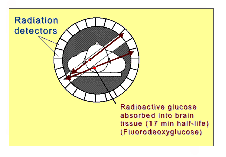
A person is injected with a slightly radioactive tracer linked to the molecule glucose (sugar). Brain (or body) tissues that are most active will use the most glucose and after a short while a lot of glucose will accumulate in active brain (or body) tissues. A by-product of radioactive decay of the tracer attached to the glucose molecule is a subatomic positron (or “antielectron”) particle which soon interacts with a nearby subatomic electron particle. This collision causes the annihilation of both particles and the emission of of two “gamma photons” which move in a straight line in opposite directions. Radiation detectors (scintillators) detect the near simultaneous arrival of the photons and, thus, can pinpoint where in the body or brain the glucose was used most heavily. [Animated image on right from https://en.wikipedia.org/wiki/File:PET-MIPS-anim.gif]

Functional Magnetic Resonance Imaging (fMRI)
Regular MRIs (see above) tell us about the structure of the brain. In the last decade, though, functional MRI (fMRI) scanners can tell us about the functioning or activity of the brain. The scanners detect where blood is being used by focusing upon the hemoglobin molecule which gives up oxygen. This is called looking for the BOLD (= Blood Oxygen Level Dependent) signal.
In an fMRI scan, a baseline structural image (MRI) of the brain is taken. Then, the brain is scanned (1) when it is not doing any task (Scan 1) and then (2) when it IS doing a specific task or activity (Scan 2). Scan 1 is subtracted from Scan 2 in order to identify those areas which are particularly active during the task.
The image to the left shows the brain as an individual is focusing upon a complex moving visual image. The areas highlighted in yellow and orange represent greater levels of activity compared to when the individual is focused upon a blank screen.
Taken from http://en.wikipedia.org/wiki/Image:FMRI.jpg
Examples of psychological findings from fMRI
- Areas of the brain which respond to pain actually do decrease their activity when taking a placebo
- Different types of memory do not activate very specific brain areas, but do activate multiple areas
- Default Mode Network (DMN): What are you doing when you are daydreaming? Imagining the future? Reminiscing about past experiences? Results from fMRI studies show that there is a specific network of brain areas which are ACTIVE when your mind is wandering in these ways. (We will discuss this more in the next class).
Head Movement: A major difficulty with fMRI imaging is the problem of head movement. Even very small motions can cause the wrong areas of the brain to display when someone is doing a task.
Limited Resolution Scale: Another major concern is the impossibility (at least now) to capture the activity of just a small number of neurons (that is, having very high and focused resolution) rather than the functions of many thousands of neurons together. Recent estimates note that cubic millimeter of cortical tissue contains anywhere from 50,000 to 100,000 neurons (e.g., see Shapson-Coe et al. 2021). Until recently, many fMRI studies could only "see" the activity of a volume of brain tissue measuring 3 cubic millimeters. That means, the joint activity of a million or more neurons being measured as a group is the best resolution scale in many past studies. Newer fMRI equipment with much higher magnetic fields are now emerging and the resolution scale is improving.
Emerging Techniques. Note that there are a host of new techniques of imaging or studying the brain which we will not review and which you will not be responsible for knowing. These include CLARITY Imaging of Tissue (Postmortem), Magnetoencephalography, Magnetic Resonance Spectroscopy, Near Infrared Spectroscopy, Syringe-Injectable Electronics, and Ultrasound Neuroimaging. For anyone in the class who expects to enter a career in neuroscience or neuro-related medicine and health care, these kinds of techniques will doubtless become more and more important in the decades ahead.
Brain Size and Intelligence
1. Humans do not have the largest brains across animal species. We do not even have the largest ratio between brain size & body size.
2. Animals with larger brains often have larger neurons so that comparing the volume of animal with human brains is not equivalent.
3. Humans: Brains vs. IQ (Intelligence)
- There is a moderate correlation between brain size and IQ tests (r = ~.30-40, which equals explaining about 9% to 16% of IQ differences due to size).
- CAUTION: IQ test scores may not be adequate to evaluate "intelligence" which is a notoriously difficult concept to quantify.
- Hevern: Intelligence involves those abilities to cope successfully within whatever environments (physical, interpersonal, or cultural) individuals find themselves.
- Because IQ tests only measure a range of cognitive functions appropriate for life in an industrialized or economically-developed world, they are not necessarily measures of the full range of coping abilities (=intelligence) that are associated with differing environments.
- Males generally do have larger brains than females (roughly 8-10%). However, there are no overall IQ differences between men and women.
- Males vs. Females
- Differences of specific skills or ability levels between men and women are sometimes found. However, within the context of differing developmental pathways (boys and girls often experience growing up with different opportunities and emphases), many such differences are not biological, but actually cultural, e.g., the superiority of boys in math may reflect the opportunities boys take to take part in activities involving numbers or geometric shapes.
- There are differences in the amount of gray vs. white matter across the sexes. Women have deeper & more sulci than men and, despite females brains being roughly 8-10% smaller in volume, there is equal cortical surface for men and women.
- Some structural differences have been found in the wiring between female and male brains. The significance of these findings is not clear.
- And, when such structural differences are found, it is not immediately clear that there is any relationship between such structural differences and actual behavioral outcome. For example, Liu et al. (2020) reported that males have a larger face processing cortical area than females; yet, studies such as Rennels & Cummings (2013) affirm that women do better than men in actual face processing tasks.
- Is there a sex/gender (s/g) difference in the human brain? Except for an overall size difference as noted above, comprehensive meta-analyses of sex/gender brain differences and their relationship to actual behavior shows very little difference. As Eliot et al. (2021) concluded their very broad review of the research literature
Despite clear behavioral differences between men and women, s/g differences in the brain are small and inconsistent, once individual brain size is accounted for. … the present synthesis indicates that such “real” or universal sex-related difference do not exist. Or at best, they are so small as to be buried under other sources of individual variance arising from countless genetic, epigenetic, and experiential factors. Thus, s/g differences in brain architecture may be similar to sex effects in gene-phenotype architecture; while statistically discernable in a very large (>100,000) sample, such effects contributed only 1.4 % to the accuracy of genotype-phenotype prediction...In layperson’s terms, these findings can be interpreted as rebutting popular discourse about the “male brain” and “female brain” as distinct organs." (p. 690)
ReferencesBlow, N. (2009, April 16). Functional neuroscience: How to get ahead in imaging. Nature, 458, 925-928. Available online at http://www.nature.com/nature/journal/v458/n7240/full/458925a.html
"Advances in magnetic resonance imaging are helping sciences learn more about the structure and function of the brain. Nathan Blow looks at how far the technology has developed and where it could go." (site blurb)
Deisseroth, K. (2015). Optogenetics: 10 years of microbial opsins in neuroscience. Nature Neuroscience, 18,(9), 1213-1225.
DeWitt, I., & Rauschecker, J. P. (2012). Phoneme and word recognition in the auditory ventral stream. PNAS, published online before print, February 1, 2012. https://doi.org/10.1073/pnas.1113427109
Eliot, L., Ahmed, A., Khan, H., & Patel, J. (2021) Dump the “dimorphism”: Comprehensive synthesis of human brain studies reveals few male-female differences beyond size. Neuroscience and Biobehavioral Reviews, 125, 667-697. https://doi.org/10.1016/j.neubiorev.2021.02.026
Liu, S., Seidlitz, J., Blumenthal, J. D., Clasen, L. S., & Razhahan, A. (2020). Intergrative structural, functional, and transcriptomic analyses of sex-biased brain organization in humans. Proceedings of the National Academy of Sciences. https://doi.org/10.1073/pnas.1919091117
Rauschecker, J. P., & Scott, S. K. (2009). Maps and streams in the auditory cortex: Nonhuman primates illuminate human speech processing. Nature Neuroscience, 12(6), 718-724. https://doi.org/10.1038/nn.2331
Rennels, J. L., & Cummings, A. J. (2013). Sex differences in facial scanning - Similarities and dissimilarities between infants and adults. International Journal of Behavioral Development, 37(2), 111-117. https://doi.org/10.1177/0165025412472411
Schmolesky, M. (2007). The primary visual cortex. Accessed February 11, 2023 from https://webvision.med.utah.edu/book/part-ix-brain-visual-areas/the-primary-visual-cortex/
Shapson-Coe, A., Januszewski, M., Berger, D. R., Pope, A., Wu, Y., Blakely, T., ... & Lichtman, J. W. (2021). A connectomic study of a petascale fragment of human cerebral cortex. BioRxiv, 2021-05. https://www.biorxiv.org/content/10.1101/2021.05.29.446289.full.pdf
This page was first posted February 3, 2005