![[Brain Image]](../graphics/head_space.gif)
PSY 340 Brain and Behavior
Class 10: Structure of the Vertebrate Nervous System
|
|
PSY 340 Brain and Behavior Class 10: Structure of the Vertebrate Nervous System |
|
Structure of the Nervous System
![[Comparison of Animal Brains]](../graphics/animal_brains_comparison.jpg)
Compare above the different types of mammal brains? What do you see?
A. Terminology to Describe the Nervous System
Central Nervous System (CNS)
- Brain
- Spinal Cord
Peripheral Nervous System (PNS)
Any nerve that does not belong to the CNS
Somatic Nervous System (SNS)
- Voluntary muscle control
- Sensory Information to the CNS
Autonomic Nervous System (ANS) • Discussed later in this lecture below
- Controls involuntary muscles & glands (heart, intestines, etc.)
- Sympathetic NS = fight or flight (or freeeze)
- Parasympathetic NS = rest & digest
Enteric Nervous System (ENS) • Often considered part of the ANS
- Controls the functions of the gastrointestinal (GI) tract -- stomach, intestines, and rectum
- Discussed in greater detail below.
Directions in the Nervous System

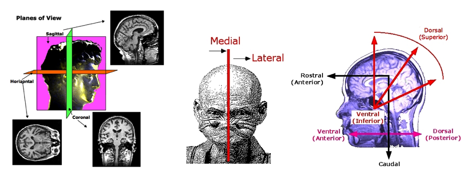
Rostral = toward the nose or front: from ROSTRUM - A bird's beak [& the prow of a ship]
- A conquering Roman admiral would address his forces from the prow of a ship captured in a sea battle. A ship's prow reminded people of a bird's beak. Hence, a rostrum became a place someone spoke from.
Dorsal = towards the back: from DORSUM - An animal or person's back
Ventral = toward the stomach or below: from VENTER - The belly or abdomen
Caudal = towards the back or the tail: from CAUDA - The tail of a horse
Superior is ABOVE and Inferior is BELOW
Proximal is NEAR while Distal is FAR
Ipsilateral occurs on the same side
Contralateral occurs on the other side
Medial is toward the middle
Lateral is toward the side
Terms about the Parts of the Nervous System
Lamina (laminae, pl.) = layer of cell bodies separated from other cell bodies by layer of axons or dendrites Column
= a set of cells aligned perpendicular to the surface of the cortex which share in the same function or properties Tract = a set of axons within the CNS which go from point A to point B. Also called a projection Nucleus (nuclei, pl.) = a cluster of neurons within the CNS Ganglion (ganglia, pl.) = a cluster of neurons outside the CNS Gyrus (gyri, pl.) = a bulge or protuberance on the surface of the cortex Sulcus (sulci, pl.) = a fold or groove which separates one gyrus from another Fissure = a deep and long sulcus
B. The Spinal Cord
- Found within the spinal column.
- Communicates with muscles and sensory organs below the level of the head
According to the Bell-Magendie law, sensory information (carried by the afferent fibers TO the brain) enters into the spinal cord by way of the dorsal roots (in the back) and motor information to the muscles and glands (carried by the efferent fibers FROM the brain) exits from the spinal cord by way of the ventral roots (in the front).
Clusters of the bodies (soma) of sensory neurons are gathered together in an area outside the spinal cord itself. The area is called a dorsal root ganglion (ganglia, plural).
The central area of the spinal cord is shaped like an "H" and consists mostly of cell bodies (or, "gray matter"). The surrounding area consists mostly of insulated axons (or, "white matter") of the spinal cord.
Damage to the spinal cord usually prevents any information from being sent to or from the brain to areas of the body controlled by the spinal cord from the level of the damage downward.
C. Autonomic Nervous System (ANS)
"Nomos" in Greek is the word for "law" or "regulation" while "autos" is the word for "self." Hence, the automomic nervous system is the set of neurons which functions almost like a law unto itself. It sends commands to and receives information from internal organs, particularly the heart, intestines, digestive system, lungs, and other organs and glands. The purpose of the ANS is to "regulate involuntary physiologic processes including heart rate, blood pressure, respiration, digestion, and sexual arousal" (Waxenbaum et al. 2022).
1. Sympathetic Nervous System (SNS)
The sympathetic branch of the ANS prepares the body for action: fight or flight. These include the heart (pumps more), lungs (breathes more), liver (releases nutrients into blood stream), stomach (stops digestion), muscles (constricts blood vessels), etc.
2. Parasympathetic Nervous System (PNS)
The parasympathetic branch of the ANS helps to restore the body, build up energy & supplies needed in the future, and relax. So, its actions include increasing digestion in the stomach and intestines, slowing down the heart, and increasing the flow of secretions such as mucus or salivation.
3. Drugs & the ANS
The two parts of the ANS tend to use different neurotransmitters,
- acetylcholine [Ach] in the parasympathetic NS
- norepinepherine in the sympathetic NS.
Note, though, Ach is also used at various stages of the SNS as well. Further research shows that there are at least 8 other neurotransmitters involved in some way in the ANS.
Many "over-the-counter" (OTC) cold and flu medications target the ANS by blocking the parasympathetic and/or stimulating the sympathetic nervous system. Sinus flow is a parasympathetic activity. The side effects of OTC remedies often relate to their pro-sympathetic system activities, i.e., increasing heart rate while drying out the mouth.
4. The GI Tract and the Enteric Nervous System (ENS)
(based on Fleming et al. 2020, Abstract)
The gastrointestinal system (GI) tract involves the stomach, small intestine, large intestine (colon), and rectum.
- The enteric nervous system (ENS), a large network of neurons that line the walls of these organs, controls how the GI tract functions. Because there are local reflex circuits, the ENS is actually capable of functioning without input from the central nervous system (CNS) although through the vagus nerve and some other connections the CNS and ENS do usually function with bidirectional signalling. The ENS has sometimes been called "the 2nd brain."
- The functions of the ENS include the propulsion of food (peristalsis) through the GI tract, as well as nutrient handling, blood flow regulation, and immunological defense.
- Increasingly researchers are finding that the microbiome (the vast number of organisms—bacteria, fungi, viruses, etc.—found in the GI tract) can have an effect on the behavior of individuals by sending signals to the CNS.
D. The Brain: Divisions
- Hindbrain
- Midbrain
- Forebrain
- Thalamus
- Hypothalamus & Pituitary Gland
- Basal Ganglia
- Basal Forebrain
- Hippocampus
Hindbrain
Medulla (medulla oblongata)
- Vital functions: breathing, heart rate, vomiting, coughing, sneezing.
- Connections through the cranial nerves (sensation & motor nerves for head) and parasympathetic nervous system.
Pons (the "bridge")
- Many nerves cross over from one side of the body to the other
- Contains (within the medulla & pons) the reticular formation (including the raphe system)
- Ascending reticular formation is also known as the ascending reticular activating system
- sends output throughout the cerebral cortex and controls our overall level of arousal and attention. It has a role in regulating many basic automatic functions (e.g., sleeping & eating)
- raphe system [pronounced "ray-fee"] • axons project widely into the forebrain. Deeply involved in our sleep-wake cycle and our readiness to respond to stimuli. These neurons form the major system using serotonin as a a neurotransmitter. Pronounced "ray-fee"
You will NOT be responsible to learn the cranial nerves.
Cerebellum ("little cerebrum" or "little brain") [Much of this is NOT in book]
Complexly connected with the cerebral cortex. We are only beginning to understand its full range of functions.
• Control of movement & balance
- Known functions with very good evidence:
• Time-related behaviors such as shifting attention for alternating sensory data (e.g., judging rhythm; playing the drums)
• Forms of simple learning & conditioning (associating stimuli with responses)
- NEW: role in cognition, language, & affect (emotion)
- The functions above have been generally assigned to two different regions of the cerebellum [see diagram on left; Marien et al. (2014)]
- Anterior: involved in motor and movement functions
- Posterior: involved in cognitive & affective (emotional) functions
- The cerebellum probably contributes to the rapid building of models of the body interacting with the world around it (= predictive brain & embodied cognition). We can therefore respond unconsciously and automatically to shifting conditions (Koziol et al. 2011)
- Most densely packed area of brain with neurons: ca. 70 billion
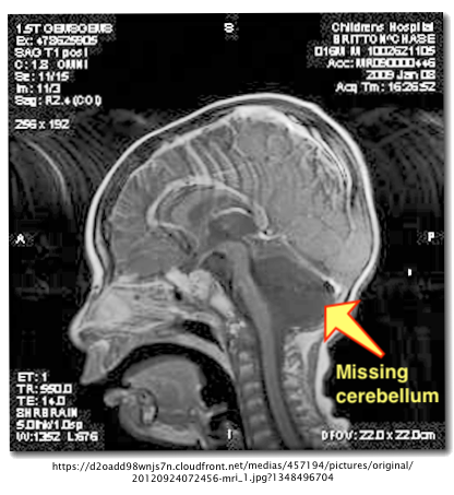
Three Strange Cases: Born without a Cerebellum (Agenesis of the Cerebellum = born without or only a partial cerebellum)
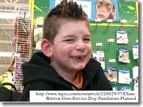
1. Chase Britton - Boy Without a Cerebellum (and Pons)


- Update on 9/17/2013: Chase is now 6 years old, has begun 1st grade (he was in kindergarten 2012-13), plays with his older brother Alex on his iPad, can count to 30, reads short words, and has a service lab/great dane dog named "Missa".
- Update on 8/14/2014: Chase attended and met with Lady Antebellum at the Erie County Fair this year.
- Updates in Dec. 2019 & May 2020. Chase is now 12 or 13 years old, uses both a wheelchair and a special walker. His father, David E. Britton, unfortunately died of stomach cancer at age 50 in May, 2020.
- 2025 Since 2020 no new updates on Chase who would now be 17-18 years old
2. Chinese Woman, 24, Found to Lack Cerebellum (2014)
In what seems to be only the 9th documented case in medical history in 2014, a 24-year-old woman in China went to the hospital complaining of a headache and was found to have been born without a cerebellum (Feng Yu et al. 2014)
3. Indian Man, 45, Found to Lack Cerebellum (2018)
- A 45-year-old man in India who had a head injury was found to lack any cerebellum in a CT scan after his accident.
- Neurologists reported that his "birth was by a normal vaginal delivery. There was no history of any difficulty in walking or in carrying out routine daily activities. The clinical and neurological examination revealed no motor impairment. No evidence of ataxia [loss of muscle control & balance], dysarthria [inability to speak clearly because of muscle weakness], nystagmus [repetitive & rapid eye movements to the side], or intention tremor [shaking of the arm/hands in reaching toward an object] was noted. The Romberg’s test [inability to maintain balance because of loss of proprioception] was negative and deep tendon reflexes were normal" (Omair et al., 2018; emphasis & descriptors within brackets [] added")
Midbrain
Tectum ("roof")
- Superior Colliculus ("upper little hill")
- Visual orientation and tracking of objects in space, e.g., a fly ball in baseball
- Inferior Colliculus ("lower little hill")
- Auditory orientation: where a sound is coming from, e.g., a car passing by a house
Tegmentum ("floor covering" or "rug")
- Ascending sensory & descending motor tracts pass through
- Several nuclei involved in the regulation of movement (e.g., eyes)
- Pain modulation
Substantia Nigra ("dark substance")
- Dopamine-producing cells which permit smooth movement of the major muscles
- Deterioration of these cells leads to Parkinson's disease
Forebrain
Outer surface = Cortex (Latin = Bark of tree) [in next class]
Limbic System (linked set of structures beneath the cortex including olfactory bulb, hypothalamus, hippocampus, amygdala, & cingulate gyrus)
- Important for behaviors related to motivation & emotion (sex, eating, drinking, fear/anxiety, & aggression)
Thalamus ("antechamber")
- sensory relay station to the cerebral cortex (except for smell)
Hypothalamus ("beneath the thalamus")
- many distinct nuclei
- regulation of motivated behaviors and internal homeostasis (e.g., temperature, thirst, hunger, sexuality)
- "Homeostatis" means that something is maintained in balance and, if it gets out of balance, it is brought back to balance.
Pituitary Gland
- beneath & connected to hypothalamus
- secretes hormones into the bloodstream (see past class)
Basal Ganglia
subcortical structures lateral to the thalamus caudate nucleus, putamen, globus pallidus multiple connections with frontal lobe cortex planning sequences of behaviors (movements), memory, emotions Basal Forebrain
- dorsal surface of forebrain
- nucleus basilis: arousal, wakefulness, attention
Hippocampus ("sea horse")
Lobes of the Cerebral Cortex - Covered in next class
- between thalamus and temporal lobe
- storage of new memories
- spatial orientation and direction of body/head
Ventricles {W} and
Cerebral Spinal Fluid (Spector et al. 2015)
The ventricles and surrounding areas of the brain are spaces that are filled with a clear fluid (cerebrospinal fluid [CSF]).
CSF is a clear, colorless, watery substance that is derived from blood plasma (to which it is similar). Normally, there are no red blood cells and very few white blood cells in the CSF. At any one time, there is about 125-150 mL (4-5 oz.) in the brain and, each day, the choroid plexus produces about 500 mL (17 oz.) of CSF which flows continuously. The CSF is reabsorbed into the blood stream by the arachnoid granulations in the subarachnoid space of the meninges at the top of the brain. (see diagram on right).
Hydrocephalus: Blockage of the flow of CSF in brain of infant causing swelling of head and, usually, intellectual deficiencies.
- Purposes of CSF: The CSF serves as a shock absorber or cushion as well as a support for the weight of the brain.
- CSF provides a range of nutrients (hormones, vitamins).
- CSF removes metabolic and other waste materials from the brain.
- Origin: CSF is formed by cells (the choroid plexus) inside the brain's ventricles
- Flow of CSF: Lateral to third to fourth ventricles. Then,
- Some CSF into the central canal of the spinal cord, while
- Other CSF flows into the space between the brain and the tissue which covers the CNS (meninges). The CSF is then reabsorbed in the subarachnoid space back into the blood.
References
Annahazi, A, & Schemann, M. (2020). The enteric nervous system: “A little brain in the gut.” Neuroforum, 26(1), 31-42. https://doi.org/10.1515/nf-2019-0027
Fleming, M. A., Ehsan, L., Moore, S. R., & Levin, D. E. (2020). The enteric nervous system and its emerging role as a therapeutic target. Gastroenterology Research and Practice, article 8024171. https://doi.org/10.1155/2020/8024171
Koziol, L. F., Budding, D. E., & Chidekel, D. (2011). From movement to thought: Executive function, embodied cognition, and the cerebellum. Cerebellum, 11, 505-525.. https://doi.org/10.1007/s12311-011-0321-y
Marien, P., Ackermann, H., Adamaszek, M., et al. (2014) Consensus paper: Language and cerebellum: An ongoing enigma. Cerebellum, 13, 386-410. https://doi.org/10.1007/s12311-013-0540-5
Omair … (2018) Primary cerebellar agenesis in a normal man. Neurology India, 66, 871-873. https://doi.org/10.4103/0028‑3886.232287
Spector, R., Snodgrass, S. R., & Johanson, C. E. (2015). A balanced view of the cerebrospinal fluid composition and functions: Focus on adult humans. Experimental Neurology, 273, 57-68. https://doi.org/10.1016/j.expneurol.2015.07.027
Yu, F., Jiang, Q., Sun, X., & Zhang, R. (2014). A new case of complete primary cerebellar agenesis - Clinical and imaging findings in a living patient. Brain, 138(6). https://doi.org/10.1093/brain/awu239
Visible Human images from <https://www.nlm.nih.gov/research/visible/visible_human.html>
Waxenbaum, J. A., Reddy, V., & Varacallo, , M. (2022, July 25). Anatomy, autonomic nervous system. In StatPerls [Internet Resource]. Treasure Island, FL: StatPearls Publishing. https://www.ncbi.nlm.nih.gov/books/NBK539845/
This page was first posted February 6, 2005