January 25. 2025
![[head]](../graphics/head.gif)
PSY 340 Brain and Behavior
Class 08: Chemical Events at the Synapse
|
January 25. 2025 |
PSY 340 Brain and Behavior Class 08: Chemical Events at the Synapse |
|
 Chemical
Activity at the Synapse: Summary
Chemical
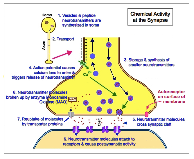
Activity at the Synapse: Summary(1) Synaptic vesicles and peptide neurotransmitters are synthesized in the soma. Vesicles are small spheres which are filled with neurotransmitter molecules (Think: a balloon blown up with water)
(2) These are transported down the interior of the axon to the presynaptic terminal (see figure below). There are molecular transporter "motors" which move the vesicles down microtubules inside the axon.
(3) Vesicles are stored and smaller neurotransmitters are synthesized in the presynaptic terminal (which is also called the synaptic bouton [or synaptic button)].
(4) Action potentials travel down the axon and cause channels to open in the presynaptic terminal. Calcium ions (Ca++) trigger the release of neurotransmitters at the presynaptic terminal membrane. This release is called exocytosis ("exo" = out of; "cyto" = cell)
(5) The molecules of neurotransmitter diffuse across the synaptic cleft (gap) to the postsynaptic membrane almost instantaneously. The molecules remain in the cleft for about 1-2 milliseconds.
- We now know that many neurons release more than one type of neurotransmitter, e.g., 2 types at the same time or one type quickly and another type slowly
(6) Neurotransmitter molecules attach to receptor sites on the post-synaptic membrane and cause post-synaptic activity (discussed below),
(7) Some neurotransmitter molecules may drift away from the cleft. Some will be destroyed in the cleft by enzymes. Others will be taken back up into the presynaptic terminal by transporter proteins (or "reptake" channels); this process is called reuptake.
(8) Back inside the presynaptic terminal, the neurotransmitter molecules may be broken up by the enzyme monoamine oxidase (MAO) or be repackaged into new vesicles when they are transported back to the soma.
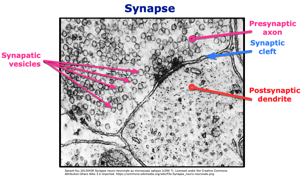
The microphotograph below shows clustered synaptic vesicles on the right of the presynaptic neuron's axon (ax), the synaptic cleft, and the postsynaptic neuron's dendrite (d). All along the presynaptic neuron membrane, the image captures synaptic vesicles merging with the membrane in the process of exocytosis. It appears online in Ackerman, Gregory, & Brodin (2012).
Here is another example of what the synaptic cleft looks like via a detailed microscopic view:
A. Types of Neurotransmitters
Type Neurotransmitters Some Important (but not all) Functions & Facts Amino Acids Glutamate (Glu) • Most widely-employed excitatory neurotransmitter in the CNS; role in long-term memory
GABA (Gamma-aminobutyric acid) • Most widely-employed inhibitory neurotransmitter int he CNS; inhibits post-synaptic neurons by hyperpolarizing them (via Cl- and K+ gates) which makes them less likely to fire.
• Effects are enhanced/increased by alcohol, sedatives, etc.
Modified Amino Acid Acetylcholine (ACh) • Skeletal motor neurons in PNS (peripheral nervous system)
• Brain: short-term memory
Monoamines Indoleamine • derived from tryptophan (an amino acid from diet). The levels of tryptophan determine the levels of serotonin. Melatonin which affects the sleep-wake cycle and we will study later is also an indoleamine.
Serotonin (5-HT or 5-hydroxytryptamine) • modulation of emotion (low levels associated with self-destructive behaviors)
Catecholamines • derived from phenylalanine (an amino acid from diet) Tyrosine
Dopa
DA/NE/Epinephrine (see below)
Dopamine (DA) • promotes coordinated muscle movement (loss = Parkinson's)
• central to experience of reinforcement (e.g., pleasure & enjoyment)
• Newer concept (not in book): tells us what we need to pay attention to: grabs our attention. Makes an experience highly "salient" or urgent.
Norepinephrine (NE) • modulation of emotion (low levels associated with depression)
Epinephrine (Adrenaline) • Stimulates sympathetic nervous system
Peptides (>100 different types)
Endorphins (& enkephalins) • suppresses transmission of pain by hyperpolarizing neurons which send out Substance P [see below]. Similar to effects of opioids (morphine, heroin, etc.)
Substance P • dull chronic pain due to nervous tissue damage or cancer ("c-fiber" pain)
Neuropeptide Y • in hypothalamus, stimulates feeding & fat storage
Purines Adenosine, ATP,
et al.• Adenosine: There are adenosine receptors in the basal forebrain cells which send signals to lead us toward sleep. Caffeine blocks these receptors and causes us to stay alert.
• ATP: basic energy molecule in all cells
Gases NO (Nitric Oxide) • dilates blood vessels & increases blood flow in brain and male sexual organ (Viagra®, Levitra®, & Cialis® affect NO functioning)
• NOT stored in vesicles, but released as needed.
• This is NOT Nitrous Oxide (N2O) which is the anesthetic "laughing gas".
Sources: Kalat (2005, 2007, 2009. 2016), Kimball (2019), Purves et al. (2001)
B. Transport & Storage of Neurotransmitter/Release and
Diffusion of Neurotransmitters
 C. Receptor Activation on the Postsynaptic Cell
What happens when neurotransmitters attach to the receptors on the postsynaptic membrane?
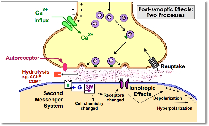
1. Ionotropic
- When neurotransmitter molecules attach to receptor, this causes the attached channel (gate) to change shape and open. These gates are called transmitter-gated or ligand-gated channels. A ligand is a chemical that binds to another chemical.
- Hyperpolarization (IPSP): opening gates to admit Cl- ions or to release K+ ions. The major neurotransmitter that causes IPSPs is GABA. The inside of the postsynaptic neuron becomes more negative.
- Effects last very briefly, ca. 5-20 mSec.
- Often found at neurons which affect vision, hearing, muscle movement, etc., i.e., neurons which respond to rapidly-changing stimuli
2. Metabotropic & Second Messenger Systems
When neurotransmitters attach to receptor, this causes a series of metabolic reactions. These reactions take a while, e.g, beginning 30 mSec or more after the release of the neurotransmitter and lasting for seconds and, even, longer:
(1) neurotransmitter molecule attaches to receptor (R in figure) which then affects
(2) a G-protein inside the neuron (G in figure). The G-protein changes shape inside the neuron and becomes activated by GTP (guanosine triphosphate). This leads to the release of
(3) a second messenger (SM in figure) such as cyclic AMP (cyclic adenosine monophosphate).
Kalat uses a metaphor which I'll borrow. Second messengers are like someone who can't get into a room, but can extend a long stick into the room. Using that stick the person releases an angry dog from a cage and that dog then runs around the room which is filled with all sorts of other animals to attack them. The dog's behavior in attacking these animals causes them to behave in a lot of different ways (e.g., to hide under or climb on top of other furniture, to defecate or urinate out of fright, etc.). Similarly, in second messenger systems, the neurotransmitter (or hormone as we will see later) never gets past the cell membrane itself, but causes something to happen inside the membrane which then affects the cell.
Second messengers can cause multiple changes inside the postsynaptic neuron, e.g.,
- open/close ion channels (add to/extend/alter ionotropic effects)
- affect the production of proteins or trigger chromosome activity.
- Second messenger effects are much slower than ionotropic effects, e.g., from 1 sec. to minutes or even hours.
Neuropeptides (endorphins, substance P, neuropeptide Y, etc.) = neuromodulators
- tend to trigger the release of more neuropeptides.
- tend to diffuse not only to postsynaptic membrane but to other neurons nearby
- often cause changes in gene activities & thus, effects can last for 20 minutes +
Variations in Receptors
- there are multiple variations of receptors to which different neurotranmitter molecules attach, e.g. ~26 types of GABA receptors, 7 families of sertonin receptors, and 5 types of dopamine receptors. Each type of receptor may have a different effect on the action of the neuron.
Drugs Binding to Receptors
- If the molecules of a drug is similar in shape to a particular neurotransmitter, that drug may be able to bind to a receptor for that neurotransmitter and, therefore, cause different kinds of effects. For example
- Nicotine, the drug found in cigarettes, stimulates a family of acetylcholine receptors call nicotinic receptors. When stimulated these nicotinic receptors tend to stimulate the release of dopamine as well. Dopamine is rewarding and, thus, nicotine feels rewarding as well.
- Opiate drugs (heroin, morphine, etc.) bind to the same receptors as the brain's endogenous morphines (i.e., endorphins) and cause effects like lowered pain, etc.
D. Inactivation and Reuptake of Neurotransmitters
- Neurotransmitter molecules may be directly taken back within the presynaptic neuron by means of transport proteins. This is called "reuptake.
- Serotonin molecules experience reuptake. Prozac and other SSRI (selective serotonin reuptake inhibitor) drugs for depression block the transport proteins (the reuptake) and cause serotonin to linger at receptors for longer periods of time.
- Similarly, methylphenidate (Ritalin), a mild stimulant used for persons with attention deficit hyperactivity disorder (ADHD), as well as cocaine block the reuptake of dopamine. However, methylphenidate works much more slowly and gradually and does not produce the "high" that cocaine does.
- Alternatively, neurotransmitters may be inactivated or broken apart (a process technically called hydrolysis) by enzyme molecules which are found in both the synaptic cleft and within the neuron.
- Acetylcholine (ACh) is used by motor neurons to signal movement. ACh is broken up by the enzyme Acetylcholinesterase (AChE) into two smaller molecules acetic acid and choline.
- Therapeutic drugs can change this. AChE inhibitor drugs can help patients with myasthenia gravis to move better and MAO Inhibitor drugs can help depressed patients feel better.
- Similarly, molecules of the monoamines (5-HT, DA, NE, epinephrine) are broken up by monoamine oxidase (MAO) found inside the presynaptic neuron
- Monoamines which are not transported back into the presynaptic neuron are broken down by the enzyme COMT (catechol-o-methyltransferase
- Note, finally, that some of the neurotransmitter molecules will simply float away from the synaptic cleft and/or be inactivated/"cleaned up" by glial astrocytes.
E. Negative Feedback from Postsynaptic Cell
- Can a neuron be told to slow down or stop releasing neurotransmitters? That is, is there some sort of a system to give the presynaptic neuron "negative feedback"? The answer is "yes". This comes about in two ways:
- Autoreceptors on the outside of the presynaptic terminal membrane: Sensitive to the amount of neurotransmitter in synaptic cleft and signals to neuron to slow/stop transmitting.
- Postsynaptic neuron can release chemicals that affect the presynaptic terminal and inhibit further release of neurotransmitters. Three chemicals have been identified: nitric oxide (NO), anandamide and 2-AG (sn-2 arachidoylglycerol). The last two bind to the same receptors as cannabinoids (the chemicals active in marijuana.)
F. Electrical Synapse (Gap Junction) {Wikipedia}
- There are a few specialized synapses in the CNS for which communication IS electrical (and are somewhat similar in function to "local neurons"). Pores on one neuron line up with pores on another neuron and form a channel (= "connexon"). Across a very very tiny gap, charged ions move easily and quickly from neuron to neuron since the channel tends to remain open all the time.
- These electrical synapses allow the body to synchronize activities which need to be done at very same time, e.g., right and left side of the lungs inhaling.
G. Hormones
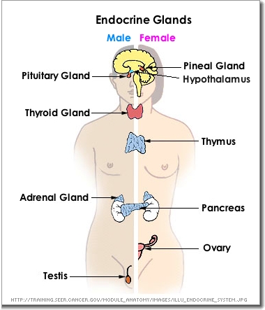
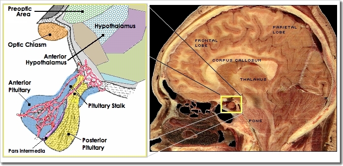
- Peptide & protein hormones will attach to the same kind of second messenger receptor sites that neurotransmitters do. However, hormones tend to have more widespread effects (compared to effects only at specific synapses).
Pituitary Gland and the Hypothalamus
- The pituitary gland has sometimes been called the "master gland" since it sends many different kinds of hormone messages after it receives instructions from the hypothalamus to which is is connected. It is comprised of two parts: Anterior pituitary and posterior pituitary (see diagram above right)
- Posterior pituitary (= an extension of the hypothalamus): made of neural tissue and secretes two hormones - oxytocin and vasopressin (or antidiuretic hormone) - which are first made in the hypothalamus.
- Anterior pituitary: made of glandular tissue that makes six different "releasing" hormones (= hormones which flow through the bloodstream and stimulate or inhibit the release of other hormones): ACTH (adrenocorticotropic hormone), TSH (thyroid stimulating hormone), prolactin, somatotropin (= "growth hormone"), FSH (follicle-stimulating hormone), and LH (luteinizing) hormone.
- We will examine the effects of these hormones later on in the course.
References
Ackerman, F., Gregory, J. A., & Brodin, L. (2012). Key events in synaptic vesicle endocytosis. In B. Ceresa (Ed.), Biochemistry, genetics, and molecular biology. doi: 10.5772/45785 Published: July 6, 2012 under CC BY 3.0 license. © The Author(s)
Kimball, J. W. (2019). Synapses. Kimball's biology pages [Online]. https://www.biology-pages.info/S/Synapses.html
Purves, D., Augustine, G. J., Fitzpatrick, D. et al (Eds.). (2001). Two major categories of neurotransmitters. Neuroscience (2nd ed.). Sunderland, MA: Sinauer Associates. https://www.ncbi.nlm.nih.gov/books/NBK10960/
The first version of this page was posted January 18, 2005.